Calcific tendonitis is a common cause of shoulder pain and is characterized by a build-up of calcium in the tendon or muscle of the rotator cuff. The Rotator Cuff is a group of four muscles and tendons that help stabilize the shoulder: Supraspinatus, Infraspinatus, Teres minor & Subscapularis. They also aid in movement: Every time you move your shoulder, you are using your rotator cuff to stabilize and help move the joint.
Calcium build-up in the rotator cuff can limit movement in the shoulder as well as cause pain.
Calcific Tendinitis – Seen on an X-Ray of the shoulder
Calcific tendonitis is a common cause of shoulder pain and is more common in people who perform a lot of overhead activities such as heavy lifting or sports like tennis.
What causes a Calcific tendinopathy?
The exact cause of calcium deposits isn’t 100% known, but there are several risk factors that predispose certain people to get them which include:
More common in women than men
People over 30 years old
Jobs or sports involving lots of overhead activity
Genetic predisposition
Metabolic disorders such as diabetes
Thyroid Disorders
Symptoms of a Calcific tendinopathy
Some patients with Calcific tendonitis may not experience pain and just complain about a limited range of motion in their shoulder. However, over time, they may start to experience pain as the condition worsens.
Diagnosis is usually made using X-ray or Ultrasound where the calcium deposit is seen in the tendon of the rotator cuff muscles.
Calcific Tendinitis
How is a Calcific tendinopathy treated?
Treatment can be conservative or surgery.
Conservative treatment can involve:
NSAIDS and painkillers
Shockwave Therapy
Therapeutic ultrasound
Rehabilitation: exercises to stabilize the shoulder
If conservative treatments do not improve the symptoms, the surgical intervention may be required.
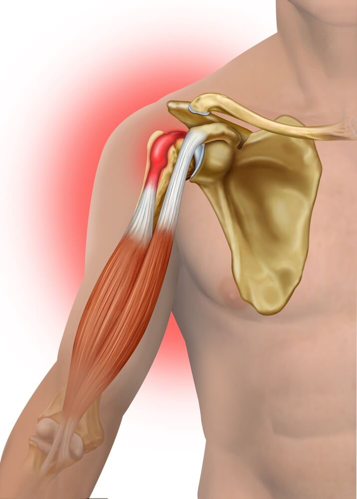
Biceps tendinopathy is a term used to describe several injuries that can occur in and around the biceps tendon:
Biceps Tendinitis – Inflammation of the tendon itself
Biceps Tendinosis – Degeneration of the tendon (non-inflamed)
Biceps Tenosynovitis – Inflammation of the tendon sheath
Biceps tendon rupture – This is usually secondary to a preexisting tear or chronic degeneration
Biceps Tendinopathy
What causes a Biceps Tendinopathy?
The exact cause of a biceps tendinopathy depends on the pre-existing factors prior to the injury as well as if it is an acute injury or results from prolonged overuse. It is also rare for a biceps tendon injury to occur on its own. It is common to find biceps tendon involvement with a rotator cuff injury, shoulder instability or labral tears. Certain sports such that involve throwing, swimming or gymnastics are also predisposing factors. As well as jobs that require a lot of overhead work.
Symptoms of a Biceps Tendinopathy
Pain in the front of the shoulder and sometimes pain into the elbow
Pain or weakness when lifting arm in front of you
Pain when raising the arm above your head, especially with activities like throwing
Possible snapping when moving the arm
Biceps tendinitis
How is a Biceps Tendinopathy treated?
Treatment for a biceps tendinopathy depends on the severity of the injury but can include:
Rest
Ice
Anti-Inflammatories or painkillers for pain management
Glenohumeral arthritis is caused by degeneration or wearing down of the cartilage layer covering the bones in the Glenohumeral joint (GHJ). This results in a bone-on-bone contact within the joint, which encourages the body to produce bone spurs, also known as osteophytes. As these rough joint surfaces rub against each other, shoulder movement becomes limited and uncomfortable. Range of motion is gradually lost as the osteophytes continue to develop.
What causes Glenohumeral Arthritis?
There are several conditions that can cause the breakdown of cartilage in a joint:
Chronic rotator cuff injury/tears: – This causes the head of the humerus to be in an abnormal position against the glenoid fossa.
Osteonecrosis: – bone death caused by loss of blood supply
Post-surgical changes that can be a result of over-tightening during instability surgery (sometimes a frozen shoulder can occur following a rotator cuff or capsule repair surgery)
Symptoms of Glenohumeral Arthritis
The two main symptoms of GHJ arthritis are Pain and Loss of range of motion:
Pain: – This may be mild to moderate in the beginning and not always there. But as the condition progresses the pain may be more persistent. Pain and stiffness experienced all day and especially at night. This can make sleep difficult at night due to it being uncomfortable to lie on one’s side.
Loss of Range of motion: – This can occur because of pain, inflammation, locking from osteophytes within the joint or from a weakness of the rotator cuff muscles (either from an old injury or surgery).
Other symptoms can include: – swelling, an abnormal resting position of the shoulder (possible hiked up), clicking or crunching felt and/or heard when moving the shoulder.
How is GHJ arthritis diagnosed?
A physical examination accompanied by diagnostic imaging (X-Ray, CT or MRI) are used to diagnose GHJ arthritis. Arthrograms are also used occasionally.
How is Glenohumeral Arthritis treated?
Conservative treatments in mild to moderate cases involve rest, NSAIDS, stretches, exercises and mobilizations of the shoulder.
More severe conditions usually require surgical intervention (either debridement or shoulder replacement).
For more information about other shoulder injuries click here
A shoulder impingement syndrome is a condition where the tendons of your rotator cuff muscles become pinched or compressed with certain movements in the shoulder. This, in turn, aggravates and damages the tendons and bursa (a fluid-filled cushion-like structure) which results in pain and inflammation. Some people with shoulder impingements find it difficult to perform daily tasks such as: putting on a seatbelt, getting dressed, raising arms above one’s head or exercising the arms (particularly overhead movements).
Prolonged impingement can lead to tendonitis or tendinosis of the rotator cuff tendons (particularly the supraspinatus) and/or bursitis (inflammation of the bursa). Calcific tendonitis (bone forming in the tendon) may also occur, and if not treated early, partial or complete tears of the supraspinatus tendon can occur, which usually requires surgical repair.
What causes an impingement syndrome?
First… some anatomy:
Place your hand on the outer top part of your shoulder and feel for a bony prominence. This bony point of the shoulder is called the acromion. The supraspinatus muscle passes underneath the acromion in the subacromial space and this is where the impingement occurs:
Now that we have a visual idea of the structures, lets explore the two different types of impingement syndromes that can occur: Primary and Secondary.
Primary Impingement Syndrome
The size of the subacromial space varies from person to person. Some people are born with a smaller subacromial space, while others may become smaller due to osteoarthritis later on in life. In both instances, the structures in the subacromial space are likely to become compressed and inflamed which results in an impingement syndrome.
Instability and dyskinesis in the shoulder can lead to improper shoulder movements. In a dynamically unstable shoulder, ligament laxity and muscle weakness can occur. The underlying cause is usually repeated overhead activity (painting or playing sports such as tennis), poor posture or from an old shoulder injury that was not rehabilated correctly.
The instability in the shoulder also results in the rotator cuff muscles working harder and eventually becoming fatigued. The rotator cuff normally functions to stabilize the glenohumeral joint but holding the head of the humerus against the glenoid fossa of the shoulder blade. When the rotator cuff begins to fail, the shoulder often hikes up which compresses the structures previously mentioned against the subacromial space.
Symptoms of an impingement syndrome
Symptoms of a shoulder can vary depending on the severity of the condition, but can include:
Pain or difficulty reaching behind your back
Pain or weakness noted when trying to lift your affected arm
Pain when lying on the affected side when sleeping
Pain radiating from the shoulder down to the elbow or hand
“Painful arc sign”
Pain when performing daily tasks such as getting dressed, putting on a seatbelt or driving.
Risk factors for an impingement syndrome
Any sport, activity or occupation that involves overhead movements will likely predispose a person to impingement syndromes. In patients older than 40 years of age, have a higher rate of incidence than males.
How is an impingement syndrome treated?
Chiropractic treatment of an impingement syndrome that does not yet require surgical repair can be conservative. This would involve mobilizations and manipulations of the affected side. Soft tissue modalities such as dry needling, massage, and stretching can also be used. Correcting the underlying instability is also essential with the above mentioned modalities as well as incorporating an exercise program to strengthen the weak muscles in and around the shoulder. Posture correction will also help to keep the shoulders in a neutral position.
The Rotator Cuff is a group of four muscles and tendons that help stabilize the shoulder: Supraspinatus, Infraspinatus, Teres minor & Subscapularis.
They also aid in movement: Every time you move your shoulder, you are using your rotator cuff to stabilize and help move the joint.
The rotator cuff is a commonly injured area and the most common injuries are strains, tendinitis, and bursitis.
What causes a Rotator Cuff injury?
There are 3 main categories when it comes to rotator cuff injuries:
Chronic Overuse injuries
Bursitis
Acute tendon/muscle tears
Chronic overuse injuries
Repeated aggravating movements over time can cause damage and inflammation to the muscles and tendons of the rotator cuff. Sports involving overhead movements such as tennis of weight training can be a culprit as well as day to day tasks or jobs such as reaching above one’s head, painting, etc. The result of repeated aggravating movements often results in a tendonosis in the shoulder. The pain can sometimes be local in and around the shoulder but pain can also be referred into the arms and hands. Poor posture also causes the shoulders to roll inwards which puts a lot of strain on the shoulder over time.
Bursitis
Bursitis is an inflammatory injury that can also occur in the shoulder. A bursa is a fluid-filled sack that sits beneath the bone and rotator cuff muscles and tendons acting as a cushion between the two structures reducing friction in the joint. The most commonly affected bursa in the shoulder is the Subacromial bursa:
Acute tendon/muscle tears
Acute injuries can occur from sports activities, falling or from prolonged overuse. The pain is usually quite severe and felt immediately after the injury occurs.
Symptoms of a rotator cuff injury
The type of pain experienced from a Rotator Cuff injury depends on the underlying cause. However, some symptoms can include:
Pain in and around the shoulder
Pain referring from the shoulder down into the arm and hand/wrist
Avoiding certain movements or activities because they cause pain (reaching above your head, lifting objects, etc)
Unable to sleep on the affected side as well as pain at night because of this
There are two risk factors to consider when it comes to shoulder injuries: Acute Traumatic injury or Chronic/degenerative injury
Acute Traumatic injury
These types of injuries commonly occur in one specific traumatic event such as lifting a heavy object falling. Acute i injuries usually occur in young people
Chronic/degenerative injury
Degenerative injuries are usually seen in older people, <40 years old. These are a result of long-term overuse and can also be associated with poor posture.
How are rotator cuff injuries treated?
Chiropractors treat shoulder injuries conservatively aiming to relieve their patient of painful symptoms as well as correcting any underlying dysfunction in the shoulder that may have caused the injury in the first place.
Treatments can include:
Mobilisation and/or manipulation of the shoulder joints
Dry needling and other soft tissue modalities
Functional rehabilitation and treatment
Taping/Strapping
Home treatments include:
Ice
Rest
Stretches and exercises
Anti-inflammatories and painkillers
If conservative treatment does not give a patient relief over several weeks to months, then surgery may be necessary.
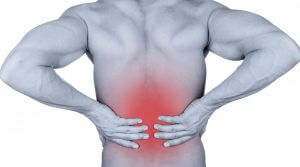
Lower back pain is very common, almost 80% of people will experience it at some point in their lives. The more common causes of lower back ache include muscle strains, acute injuries, overuse or specific conditions such as:
The symptoms of lower back pain can vary depending on the underlying condition or injury. Well break it down into 3 categories:
Referred Pain, Sciatica
Localised Pain
Muscle Pain
Referred pain
Pain shooting down the legs is usually called sciatica. Disc herniations or spinal stenosis commonly present as an electric or shooting pain referring down the thigh and leg. This is because of a compression of the nerve roots as they exit the spine. Acute facet syndromes (which patients often call a “pinched nerve”) can also cause pain referred into the butt and lower limb.
Facet Joint Pain Referral Patterns
Localised pain
In cases of muscle strains, facet dysfunction or degeneration, a person may experience pain in and around the lower back. This is due to inflammation and aggravation of the joints of the spine
Localised Lower Back Pain
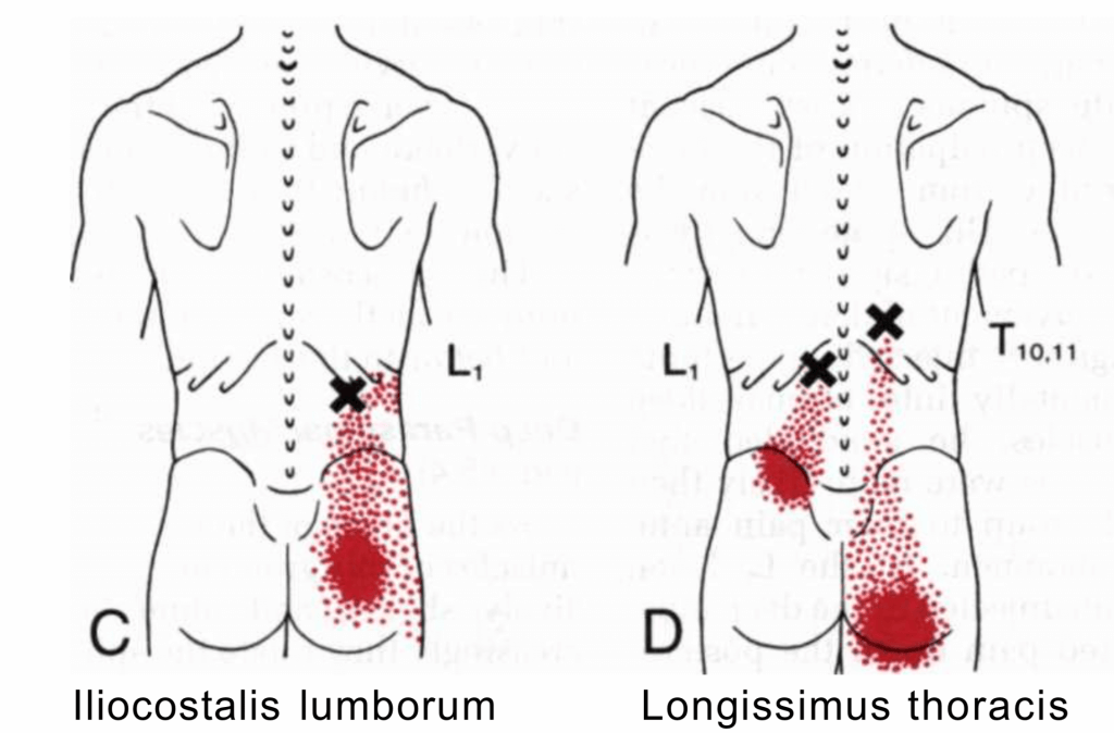
Muscle pain
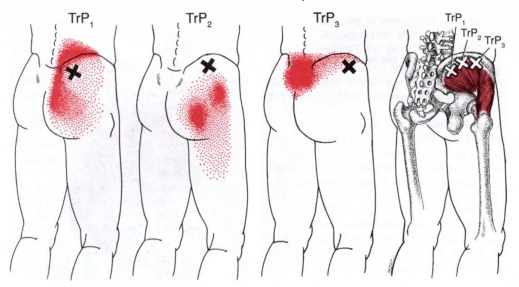
In almost all cases of lower back pain, there will be muscle involvement. This may present as a person simply experiencing localised pain in the lower back. However, muscles can also refer pain into the butt or lower limb. This type of pain referral is commonly seen in cases of myofascial trigger points.
Trigger point referral of lower back some musclesTrigger point pain referral from the gluteus medius to the lower backGluteus Minimus Trigger point referral – Often called Pseudo Sciatica
Treatment for lower back pain
Chiropractors may utilise several treatment methods and modalities to treat lower back pain:
Spinal manipulation/Adjustments
The technique most commonly used by chiropractors is the adjustment. This helps to relieve lower back pain by reducing pressure on sensitive structures such as nerves. It also increases movement and flexibility, increases blood flow and reduces muscle tension.
Lower back adjustment
Decompression techniques
These include lumbar traction or flexion-distraction. Our spines are constantly under compressive forces and applying a traction force greatly helps offload the joints and discs within the spine. Inversion tables can also be used for decompression of the lower back.
Inversion table
Soft tissue therapy
When a problem occurs in the lower back, a common response in the body is a reactive muscle spasm to try and protect an aggravated area of the spine. Massage, fascial release and instrument-assisted soft tissue mobilisation (IASTM) are very effective in relieving muscle tightness in lower back pain.
When muscles become tight, it is possible to feel a knot or a tight band within a muscle. These knots are called trigger points and are often treated using dry needling. This involves the practitioner inserting an acupuncture needle into the muscle, which stimulates blood flow to the affected area and results in a reduction of pain, inflammation and muscle tension.
Dry Needling
Other modalities
Heat and cryotherapy can assist in pain relief and decrease inflammation. Electrotherapy such as interferential current or ultrasound may also be used.
Ultrasound
IFC
Exercise and ergonomics
One of the most common underlying causes of lower back pain is poor posture and weak core muscles. Incorporating a good exercise program as well as changing work habits, altering desk arrangements and making sure that you are sitting correctly is extremely important as these will help maintain the positive effects of your back pain treatment.
One of the most common questions my patients ask me is "What is the cracking sound during an adjustment?" or "Is something breaking??"
This sound is known as a Cavitation or an audible release. It is caused by the opening of the joint surfaces which takes place during an adjustment. Each joint contains a liquid called synovial fluid which reduces friction between the joint surfaces. When the joint surfaces are opened, the pressure in the joint capsule is lowered which allows for an expansion of gas bubbles in the synovial fluid. These bubbles persist for approximately 30 minutes before the gas is absorbed back into solution.
Like back pain, neck pain is very common, we\’ve all experienced it at some point in our lives.
Some common causes of neck pain can include muscle strains, acute injuries such as whiplash from a motor vehicle accident, poor posture and bad work/desk ergonomics. Other causes of neck pain can include:
Neck aches and pains from sitting in front of a computer all day (which I am experiencing right now 😅)
Symptoms of neck pain
The symptoms of neck pain can vary depending on the underlying condition or injury. Well break it down into 3 categories:
Localised Pain
Muscle Pain/tightness
Referred Pain
Localised pain
In cases of muscle strains, facet dysfunction or degeneration, a person may experience pain in and around the neck. Neck pain can be experienced in the front (anterior) as well as at the back (posterior) of the neck. The pain is usually due to inflammation and aggravation of the joints, ligaments, intervertebral discs and muscles of the neck.
Muscle pain
When the muscles in the neck and shoulders become tight or inflamed, they can produce painful symptoms. These symptoms can be local to the affected area or they can refer pain as seen with myofascial trigger points. Trigger points are taut bands or \”knots\” that can be felt in the muscles. Trigger points refer pain in a predictable pattern, this can be pain referred around the neck and shoulders, to the head, down the back or into the arms and hands. Here are some examples:
Trapezius Muscle
Levator Scapulae Muscle
Scalene Muscles
Sternocleidomastoid (SCM) Muscle
Referred pain
There are different kinds of referred pain that a person can experience, we have already covered the pain referral patterns of specific muscles above. A shooting, burning or electric shock-like pain felt going down the arm and sometimes into the arm is called radicular pain. Radicular pain occurs in cases of spinal stenosis, disc herniation with nerve root compression, cervical facet joint syndromes or degeneration, as well as thoracic outlet syndrome. Pins & needles, tingling, numbness and a loss of grip strength can also occur.
Treatment for neck pain
Chiropractors may utilise several treatment methods and modalities to treat neck pain:
Spinal manipulation/Adjustments
Adjusting/manipulating the neck helps to relieve pain by reducing pressure on sensitive structures such as nerves, increases movement and flexibility, increases blood flow and reduces muscle tension. When an adjustment takes place, normal nerve function is restored to the joint and very often you will find that a muscle spasm that was previously there has subsided almost instantly.
Decompression techniques
Manual traction or door-traction units can be used to help relieve neck pain and radicular symptoms. Our spines are under constant pressure so these types of treatment modalities are extremely useful in treating neck pain.
Soft tissue therapy
A common response to pain in the body is a reactive muscle spasm to try and protect an aggravated area of the spine. Massage, fascial release and instrument-assisted soft tissue mobilisation (IASTM) are very effective in relieving muscle tightness.
Dry needling
A very effective method for treating myofascial trigger points is Dry Needling. This involves the practitioner inserting an acupuncture needle into the muscle, which stimulates blood flow to the affected area and results in a reduction of pain, inflammation and muscle tension.
Other modalities
Heat and cryotherapy can assist in pain relief and decrease inflammation. Electrotherapy such as interferential current or ultrasound may also be used.
Exercise, Stretching and Ergonomics
A common pattern of muscle imbalance seen in the neck is something known as Upper Crossed syndrome. Here we\’ll find that a person has tight upper trapezius, levator scapulae and pectoralis manor & minor muscles, with weak deep neck flexors, rhomboid, serratus posterior and lower trapezius muscles. When this occurs, it\’s common to see a person has a rounded shoulders, hunched forward and with their head in the forward position. This also just happens to be the position most us are sitting in while we\’re in front of a computer.
To correct this there are a few factors:
Strengthen the weak muscles
Stretch the tight muscles
Make sure you are sitting correctly (see pictures below)
Take regular breaks while working at your desk to stand up and walk around. Over time we tend to slouch while sitting so it\’s important to reposition yourself regularly.
A Disc Herniation, sometimes called a Slipped Disc, is an injury which occurs in the neck or back where part of the intervertebral disc bulges out and presses on a nerve in the spine. One of the most common symptoms of a disc herniation is a shooting type of pain going down into the arms or legs. This type of pain is typically referred to as radicular pain.
In addition to the typical shooting pain, it is also possible to experience localized pain around the spine pain from the disc itself, muscle spasms, as well as active myofascial trigger points in the muscles around the affected area. Muscle weakness, loss of sensation or even muscle atrophy as a result of prolonged nerve root compression can also occur.
Things that typically aggravate a disc herniation can include: bending or leaning forward, standing, walking or sitting for long periods and pain while sneezing, coughing or straining while on the toilet.
Usually, a clinical examination will reveal positive nerve root tension tests and imaging studies such as CT or MRI will show that part of the intervertebral disc (IVD) is bulging out and pressing on a nerve root. We’ll specifically be looking at the lumbar spine as an example, but disc herniations can occur throughout the spine.
What are the potential causes of a disc herniation?
Disc herniations can occur from some of the following:
Chronic/prolonged overloading of the disc, usually from activities that involve heavy lifting.
A previous injury to the lower back
Bad posture, muscle imbalances and congenital defects in the spine structure.
Age-related degeneration
The above causes are also dependent on certain risk factors such as:
Genetics
Weight (obesity in particular)
Systemic conditions (e.g. rheumatoid arthritis)
Lack of exercise and weak core muscles
An increased curve in the lumbar spine/hyperlordosis from an Anterior pelvic tilt.
Anatomy
Let's explore the anatomy of the spine to help better understand how and why a disc herniation occurs. This is where my amazing drawing skills come into play ?
Each vertebra in the spine has an anatomical cushion or shock absorber between each vertebral body, the IVD. The size of the IVD corresponds to the vertebra it sits in-between; They start off fairly small in size at the top of the cervical spine and increase in size moving down through the thoracic spine and to the lumbar spine as the weight of the trunk increases.
The IVD is made up of 2 structures:
1.Annulus Fibrosis - An outer layer of fibrocartilage rings
2.Nucleus pulposus - An inner gel-like centre
Figure 1: Anatomy of an intervertebral disc
The 2 parts of the IVD allow for even weight distribution at each vertebral level. This, however, changes when there is damage or degeneration in the IVD occurs.
Disc degeneration and the stages of disc herniations
Degeneration... Scary word right? ? Well, the truth is that osteoarthritis is a very common occurrence from years of wear and tear on the cartilage and joints. In the spine, the IVD's can become dehydrated and start to thin out, which reduces its shock absorbing ability. There are some predisposing factors that will obviously speed up the process such as those mentioned earlier, but in general, the nucleus of the intervertebral disc becomes dehydrated as a normal part of aging just like any other cartilaginous joint in the body.
Degeneration in the spine usually involves narrowing or flattening of the IVD’s and wearing down of the cartilage in the facet joints. Over time these joint surfaces become rough and bumpy with additional bone growths such as osteophyte formation will occur.
Figure 2: Stages of degeneration in the lumbar spine
Stage 1: Degeneration
The fibrocartilage rings of the annulus are susceptible to tearing as a result of an injury or age-related degeneration. These tears can occur in two directions: circumferentially or radially.
Circumferential tears are believed to be more common and indicate a separation of the layers of concentric fibrocartilage of the annulus.
Radial tears usually start from the inner part of the annulus (near the nucleus) and progress outwards.
Once the tearing has occurred, nerve endings within the annulus the become extremely sensitive to inflammation and pressure, which will result in a lot more pain sensation compared to other/healthy discs in the spine. One unfortunate trait of the IVD is that only the outer 1/3 of the annulus is innervated by nerves (in a healthy disc). So a patient could possibly have tearing in the inner annulus and not experience any painful symptoms until it becomes quite advanced. The irony of this is that once a disc has become damaged, more nerve receptors, specifically ones that detect pain, develop deeper into the layers of the annulus.
Stage 2: Prolapse
The nucleus then starts squeezing through the space in the annulus until it eventually starts to bulge outwards:
Stage 3: Extrusion
If the bulge progresses, it will eventually herniate out of the disc and can then put pressure onto a spinal nerve root.
At this point we can consider that a patient may be experiencing pain from one source or a combination of different sources:
From the direct pressure of the nucleus onto a nerve root causing the typical shooting pain down the leg, also known as Sciatica,
Pain arising from the disc,
Pain due to irritation of the nerve roots as a result of the inflammation response to the nucleus protruding into the vertebral canal.
Stage 4: Sequestration
The next stage of disc degeneration doesn’t always occur but it is still a possibility and that is Sequestration of the herniating nucleus. Here part of the herniating nucleus will break off and cause all sorts of trouble as it's a loose body within the spinal column:
It is also possible that the disc bulge can be larger, occur on the opposite side or centrally compressing the spinal cord:
Why does bending forward hurt so much?
Certain movements will cause a lot of pain if a person has a disc herniation, specifically leaning or bending forward into a flexed position. This occurs because of the biomechanics involved in this type of movement. Usually, when you flex forward, the top vertebra slides forward on top of the vertebra below. This causes compression and wedging of the IVD in the front, which in turn causes the nucleus to be pushed backward. In a healthy disc, the nucleus stays within the confinements of the disc, but if the annulus is torn from injury or degeneration, it will bulge out and compress the spinal cord and/or nerves. This emphasizes the importance of squatting down and using your legs to lift heavy objects.
So, how do you treat this?
Well there are 2 main options:
Conservative treatment
Surgery - laminectomy or disc replacement.
Chiropractors will usually treat disc herniations conservatively using the following treatment techniques:
Spinal decompression in the form of flexion distraction or lumbar traction,
Adjusting the lumbar spine
Soft tissue therapy such as dry needling, massage, and fascial release
Recommending exercises and stretches to help strengthen the core musculature and help to correct any postural imbalances that the patient may have.
Better ergonomics, especially if you're sitting at a desk all day
Can the IVD heal on its own over time?
The IVD is similar to cartilage, being that is has a very limited blood supply and this slows down the healing process quite a lot. A healthy IVD won't spontaneously herniate so there must be something that is putting that disc under a lot of pressure that makes it susceptible to injury. This is why finding the underlying cause is a very important part of the treatment instead of just treating the symptoms.
If you have any questions about disc herniations, back pain or would like to book an appointment, please feel free to contact me here